
X-Ray Machines: Essential Imaging Technology for Fast and Accurate Diagnosis
In modern healthcare, medical imaging plays a crucial role in identifying diseases, injuries, and internal abnormalities at an early stage. Among the most widely used and reliable diagnostic tools is the X-ray machine, a fundamental imaging system that uses controlled radiation to produce clear images of bones, organs, and other internal body structures. Because of its speed, accuracy, and accessibility, X-ray technology remains one of the first-line diagnostic methods in hospitals, clinics, and emergency care centers worldwide.
X-ray examinations are commonly performed to detect bone fractures, joint dislocations, chest infections, lung diseases, dental problems, and certain tumors. Physicians frequently rely on chest X-rays to evaluate conditions such as pneumonia, tuberculosis, and heart enlargement, while orthopedic specialists use skeletal X-rays to assess trauma-related injuries. The ability to obtain immediate visual information makes X-ray imaging essential for rapid clinical decision-making and timely treatment.
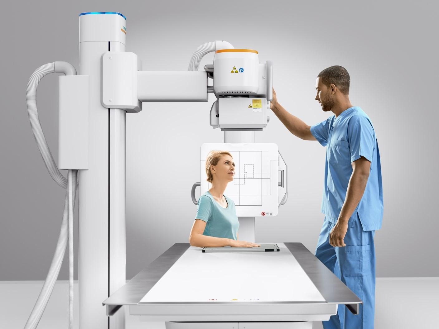
The X-ray procedure is quick, painless, and non-invasive. During the examination, the patient is positioned between the X-ray source and a digital detector or film plate. A brief burst of radiation passes through the body, and different tissues absorb the rays at varying levels. Dense structures like bones appear white, while softer tissues appear in shades of gray, producing a clear diagnostic image within seconds. Most X-ray scans take only a few minutes, allowing efficient patient flow in busy medical environments.
One of the major advantages of modern X-ray machines is the transition from traditional film to digital radiography. Digital X-ray systems provide higher image quality, faster image processing, lower radiation exposure, and easy electronic storage for future comparison. Advanced features such as portable X-ray units, mobile radiography, and real-time image sharing further enhance clinical efficiency, especially in emergency rooms, intensive care units, and remote healthcare settings.
X-ray imaging is also valuable for monitoring treatment progress and postoperative recovery. Doctors use follow-up X-rays to evaluate bone healing, check implant positioning, and observe changes in lung or abdominal conditions. This continuous imaging capability supports better long-term patient management and improved medical outcomes.
Although X-ray examinations involve a small amount of ionizing radiation, the exposure level is carefully controlled to ensure patient safety. Modern equipment is designed to use the lowest effective radiation dose while still producing high-quality diagnostic images. When medically justified, the benefits of accurate and rapid diagnosis significantly outweigh the minimal associated risks.
Like all medical imaging techniques, X-ray machines have certain limitations. They may not provide sufficient detail for soft tissues compared to CT or MRI scans, and additional imaging tests may sometimes be required for comprehensive evaluation. Nevertheless, due to their affordability, speed, and diagnostic value, X-ray systems continue to be one of the most essential tools in everyday medical practice.
In conclusion, the X-ray machine remains a cornerstone of modern diagnostic medicine. Its ability to deliver fast, reliable, and cost-effective imaging supports early disease detection, emergency assessment, and ongoing treatment monitoring. As digital technology continues to advance, X-ray systems will remain indispensable for improving healthcare quality and patient outcomes across the world.

