
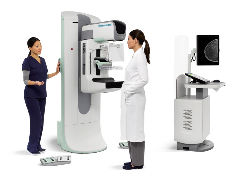
Mammography Systems: Advanced Imaging for Early Breast Cancer Detection
In modern healthcare, early detection plays a vital role in successfully treating life-threatening diseases. Among the most effective technologies for women’s health screening is the mammography system, a specialized medical imaging device designed to examine breast tissue in detail. By using low-dose X-rays to produce high-resolution images, mammography enables healthcare professionals to detect breast abnormalities at an early stage, often before physical symptoms appear.
Mammography is widely recommended as a routine screening tool for women, particularly those over the age of forty or individuals with a family history of breast cancer. The technology helps identify tiny tumors, calcifications, cysts, and other structural changes within breast tissue. Detecting these conditions early significantly increases the chances of successful treatment, reduces mortality rates, and improves long-term patient outcomes.
The mammography procedure is quick, safe, and minimally uncomfortable. During the examination, the breast is gently positioned and compressed between two plates to obtain clear and accurate images. This compression is essential for reducing motion, improving image clarity, and minimizing radiation exposure. The entire screening process usually takes only a few minutes, allowing patients to resume normal activities immediately afterward.
One of the most important advantages of modern mammography systems is their ability to provide high-resolution digital imaging with enhanced contrast and detail. Advanced technologies such as digital mammography, 3D tomosynthesis, and computer-aided detection support radiologists in identifying suspicious areas more accurately. These innovations reduce false negatives, improve diagnostic confidence, and enable earlier medical intervention when necessary.
Mammography systems are used not only for routine screening but also for diagnostic evaluation when symptoms such as breast lumps, pain, nipple discharge, or skin changes are present. In addition, they guide biopsy procedures and help monitor treatment progress in patients already diagnosed with breast disease. This makes mammography an essential component of comprehensive breast care services in hospitals, cancer centers, and diagnostic clinics.
Although mammography involves a small amount of radiation, the dose is carefully controlled and considered safe for medical screening. The clinical benefits of early cancer detection far outweigh the minimal risk associated with exposure. Continuous technological improvements have further reduced radiation levels while enhancing image quality and patient comfort.
Like any medical examination, mammography may have certain limitations. Dense breast tissue in younger women can sometimes make interpretation more challenging, which is why additional imaging methods such as ultrasound or MRI may be recommended in specific cases. Nevertheless, mammography remains the gold standard for breast cancer screening worldwide.
In conclusion, the mammography system is a crucial technology for protecting women’s health through early detection and accurate diagnosis of breast disease. Its ability to reveal hidden abnormalities, support timely treatment, and improve survival rates makes it indispensable in modern medical practice. As awareness of preventive healthcare continues to grow, mammography will remain a cornerstone of effective breast cancer screening and women’s healthcare services.

